Experimental and neuropathological evidence continues to position the vagus nerve as a central conduit in Parkinson’s disease and as a modulatory node in Alzheimer’s disease. Animal models and human tissue studies link retrograde transport of misfolded α‑synuclein from the gut to the dorsal motor nucleus of the vagus (DMnX) in the brainstem. Neuropathologists describe the DMnX as “a primary site of pathological α‑synuclein deposition,” according to Ruth E. Musgrove et al.
In experimental work, excess α‑synuclein reduced DMV neuron surface area by about 42% (from ~3,800 μm² to ~2,200 μm²) and altered ion-channel activity that can slow neuronal pacemaking. Clinical imaging and ultrasound studies report modest bilateral atrophy of the vagus nerve in Parkinson’s patients. Epidemiological signals—such as lower Parkinson’s rates after complete truncal vagotomy in some cohorts—support, but do not prove, a gut‑to‑brain route in a subset of cases.
Alzheimer’s links are less about a single protein moving along the nerve and more about network and inflammatory effects. Degeneration in basal forebrain cholinergic neurons, locus coeruleus pathology and impaired cholinergic anti‑inflammatory signaling intersect with vagal pathways. Ruth Narramore of Sheffield Teaching Hospitals notes that “the accumulation of amyloid and tau in the LC is an early pathological feature preceding clinically detectable memory impairment by decades.” Disrupted vagal regulation can amplify neuroinflammation and HPA‑axis changes that harm hippocampal circuits.
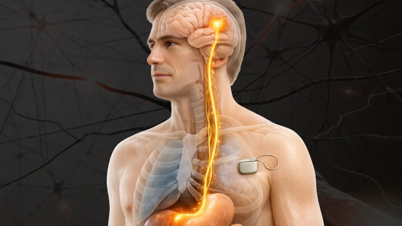
Vagus nerve stimulation (VNS), including noninvasive transcutaneous approaches (taVNS), is being tested as a way to boost neurotransmitter release and reduce inflammation. A 2025 meta‑analysis of six randomized trials (176 participants) reported that tVNS improved motor severity scores and gait during on‑medication states (standardized mean difference −0.48). A 2022 double‑blind trial of taVNS in mild cognitive impairment found a 3.2‑point MoCA‑B gain versus 0.3 points for sham (p = 0.033) over six months. Authors and reviewers caution that sample sizes, stimulus protocols and outcome measures remain inconsistent across studies.
Researchers call for larger, standardized trials with objective biomarkers (fMRI, EEG, VSEP) and clearer stimulation parameters to test whether VNS can alter disease course. Qian Hu and colleagues summarize the hypothesis: “VNS likely exerts neuroprotective effects through multiple convergent pathways, including neuromodulation, anti‑inflammatory effects, the reduction of oxidative stress, neurotransmitter regulation, and functional brain network reorganization.”
Educational and anatomical work remains important for training clinicians and researchers. Cadaver dissection and pathological specimens help students trace vagal pathways and early brainstem nuclei involvement. The Institute of Human Anatomy, for example, uses cadaver‑based resources alongside digital tools to illustrate how vagal pathology maps onto early autonomic symptoms such as constipation.
Overall, the vagus nerve is a repeat focus in current research: a possible route for α‑synuclein in Parkinson’s, a regulator of cholinergic and inflammatory cascades in Alzheimer’s, and a target for neuromodulation trials that are promising but not yet definitive.
Photo credit: kajabi-storefronts-production.kajabi-cdn.com
Tags: vagus nerve, Parkinson's disease, Alzheimer's disease, vagus nerve stimulation, taVNS
Topics: Vagus nerve & taVNS, Neuromodulation, Non-invasive brain stimulation